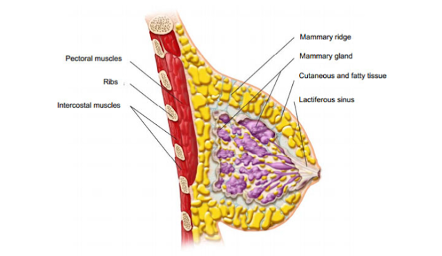
Your mammary gland, known to the layman as the breast, has a relevance that really needs no introduction. Following the womb, it is the organ with which a mother ensures the survival of her child. It offers the first food every human gulps down with a reflex hardwired within our system. In the subsequent lines we will discuss the structure of the human breast in great detail, and so the viewers can acquire sufficient biological knowledge. This will not only help you take better proper care of this treasured part of your body, but also be alert to capture if there's something incorrect.
BREAST AND SURROUNDING BUILDINGS
The human breast is actually an evolved form of the modified skin appendage, the sweat glands. They are not so developed in the males but undergo a lifetime of development and changes in the females, particularly during the reproductive system amount of life. A match of mammary glands snooze on the upper upper body wall on a muscle called the pectoralis major, as shown in the diagram. Beneath this muscle lies the pectoralis small, which in turn is placed on the chest wall membrane made up of the ribs and intercostal muscles. The breast growth is surrounded and maintained upper chest fat pad. The appendage is covered by skin area while at the centre is a pointed framework called the nipple, from where the milk ejects out. The nipple is surrounded by a dark brown pigmented area called the areola which contains easy muscle. This area is between long coarse frizzy hair. How big the breast differs widely, the appropriate size being subjective and affected by cultural norms.
THE BREAST TISSUE
To put it simply, the breast tissue is consisting of two parts: the parenchyma and the stroma.
The parenchyma: this is the key glandular tissue of the breast. To have an improved understanding, consider the diagram to visualize the main points being presented. This part commences at the left nip where six to five major ductal systems begin. The keratinizing epithelium of the skin follows into these ducts and a keratin plug is often available at their orifice. This kind of is then dilated part of the ducts understands as lactiferous sinuses. Below the milk accumulates before it is released. Following this, successive branching of the bigger ducts takes place into smaller ones. At some point, these smaller ducts open up into precisely what is called the Terminal duct lobular unit (TDLU). Every single unit is composed of a terminal duct branching off into small grape like structures called the acini, that are in charge of the production of milk. Every ductal system occupies one quadrant of the breasts, which extensively overlaps each other. The terminal ductwork and lobules in the standard breast are composed of two styles of cells. A single that is towards the lumen produces milk. The other lies beneath it and is composed of contractile cells that help in ejection of dairy during lactation.
The stroma: this is the encouraging tissue of the breasts. It is basically a blend of dense fibrous combinatorial tissue and adipose tissues . Younger women have more dense tissue and as they get old, it is replaced by more and more embonpoint tissue rendering it more radiolucent on mammograms. This makes any sort of unusual density better to catch. Encircling the lobules is a stroma that is receptive to hormones, giving go up to the alterations you feel during your monthly menstrual period.


